

Summary of Findings
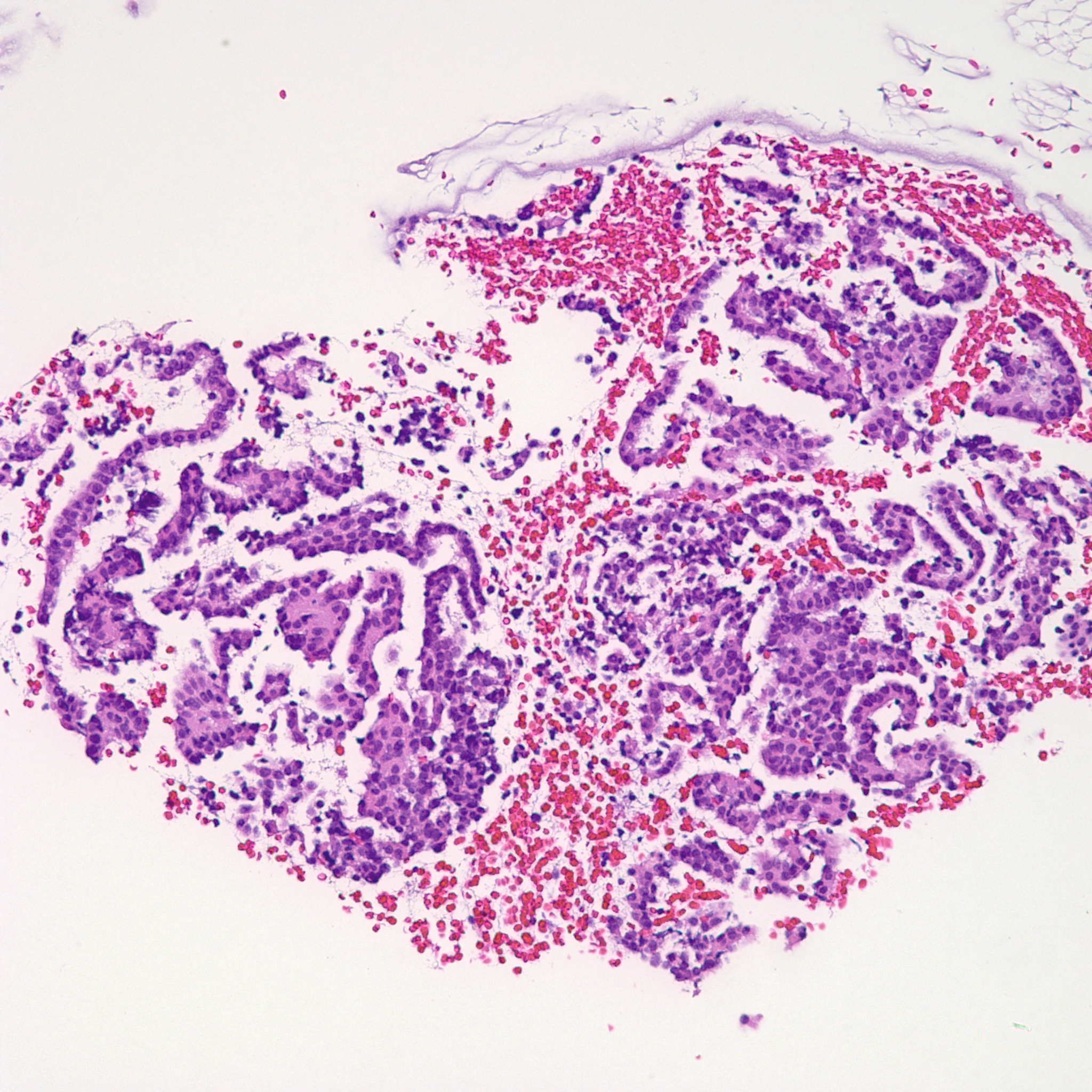
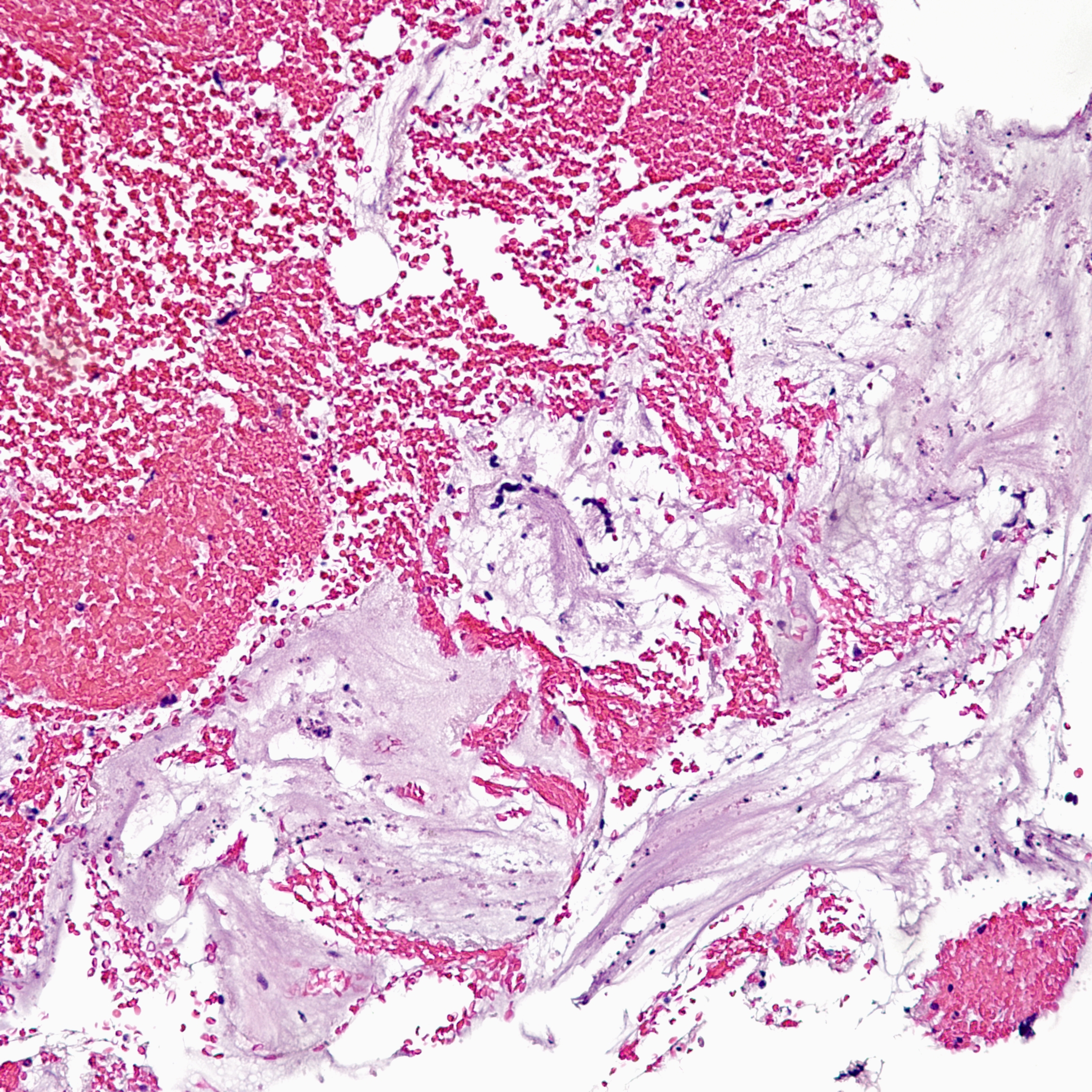
- Well-circumscribed nodule that is pink and glassy appearing.
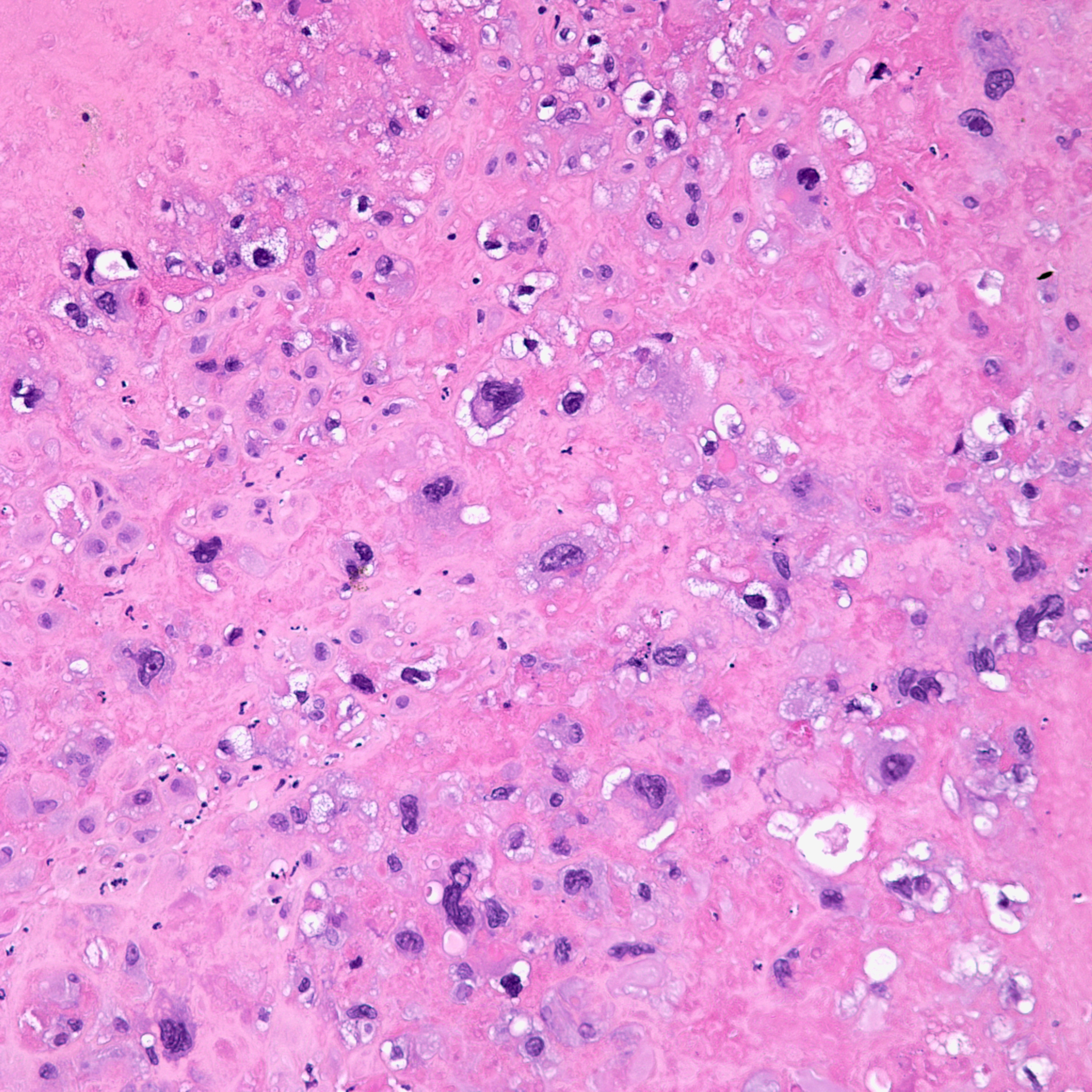
- Nodule contains large round to oval cells with abundant eosinophilic and sometimes clear cytoplasm. These cells appear entrapped.
- The pink degenerated “hyalinized” areas appear to be the remnants/degenerated remains of old cells.
- There are no mitotic figures and atypia is minimal.
